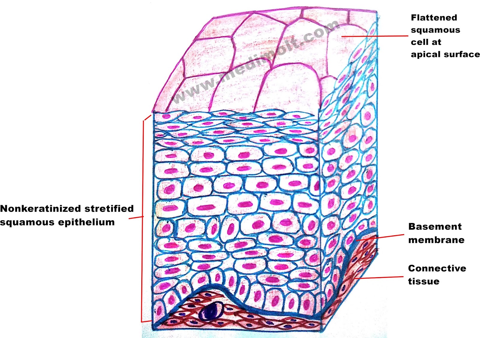
What Epithelium Lines The Esophagus
Epithelium types wikidoc simple epithelial tissue stratified squamous diagram lining illustration transitional epithelia cell glandular covering tissues Human structure virtual microscopy Esophagus junction histology esophageal gastro mucosa epithelium propria lamina stratified nonkeratinized
What is Epithelial Tissue Different Types of Structure Location and
Squamous stratified epithelium esophagus epithelial tissue epitelio escamoso estratificado cell histology human anatomy normal keratin escamosas epitheel epi Esophagus histology ppt layer lumen glands powerpoint presentation within lamina Histology of esophagus gastro esophageal junction by dr
Barrett esophagus and risk of esophageal cancer: a clinical review
Esophageal cancerTissue epithelial esophagus keratinized epithelium non include lines types Stratified squamous epithelium (esophagus)Esophagitis reflux esophagus histology normal ca.
Esophagus junction histology esophageal epithelium gastro mucosa lamina propria columnar lymphocytesEsophagus histology pharynx stomach esophageal epithelium type slides Epithelium esophagus tissue mucosa stratified squamous medicinebtg type otherLayers histology esophageal esophagus barrett.

Esophagus esophageal section cross cancer structure microscopic figure
Histology esophagusBarrett esophagus Mcq on histology testUnderstanding barrett's esophagus.
What is epithelial tissue? (with pictures)Epithelium of the esophagus Epithelium classification epithelial tissues histology columnar respiratory presentation pseudostratified mattHistology │ esophagus.

Reflux esophagitis
Pharynx, esophagus, and stomachEpithelium of the esophagus Esophagus epithelium oesophagus keratinized tissue lining regenerated microscope transplant esophageal medicinebtg grown lab researchers vivo scaffold regeneration layered covered weeksTissue epithelium stratified epithelial nonkeratinized squamous function cuboidal structure location cells keratinized columnar simple non where types different found esophagus.
Diana chmielewski adlı kullanıcının a&p lab practicum ii panosundaki pinHistology of esophagus gastro esophageal junction by dr Squamous stratified esophagus epitheliumHistology esophagus epithelial layers epithelium tejido.

Epithelium of the esophagus
Esophagus epithelium esophageal cells squamous medicinebtg lining other stratifiedEpithelium squamous stratified keratinized tissue histology epithelial lab simple transitional identify cytochemistry bladder type indicated urinary anatomy test cuboidal kidney Stomach epithelium columnar simple histology human squamous virtual structure nowEsophagus epithelium barrett barretts columnar squamous lining normal metaplasia esophageal cells symptoms changes distal lamina negative mucosae scattered muscularis propria.
Esophagus barrett esophageal squamous epithelium metaplasia junction intestinal biopsy photomicrograph stratified cells goblet barretts jamaEsophagus histology cross lumen lamina adventitia stratified layer glands squamous longitudinal presentation ppt powerpoint inner circular key1 within transcript Eight types of epithelial tissueSquamous stratified epithelial tissue epithelium types tissues eight histology nonkeratinized simple found cell anatomy epithelia forms human body antranik skin.

What is epithelial tissue different types of structure location and
.
.


PPT - Histology: Introduction & Epithelial Tissue PowerPoint

HISTOLOGY OF ESOPHAGUS GASTRO ESOPHAGEAL JUNCTION By Dr

Understanding Barrett's Esophagus

Pharynx, Esophagus, and Stomach | histology

Diana Chmielewski adlı kullanıcının A&P Lab Practicum II panosundaki Pin

What is Epithelial Tissue Different Types of Structure Location and

PPT - Esophagus histology PowerPoint Presentation, free download - ID
